Microscope Slide Specimen - Sample Images
These are sample photos of prepared microscope slide specimens. Enlarged images were captured through a microscope using our DSLR Microscope Adapter or compact digital camera adapter, then resized for website display.
PREPARED SLIDES
BIOLOGICAL MICROSCOPY
EDUCATIONAL SPECIMENS
WHOLE ORGAN SLIDES
About These Microscope Specimen Images
The images below are reference examples for prepared slide observation and microscope photography. Image appearance may vary depending on the microscope, objective lens, illumination method, camera adapter, exposure settings and specimen condition.
Click each image to view a larger image.
Educational Purpose Specimen - Sample Images
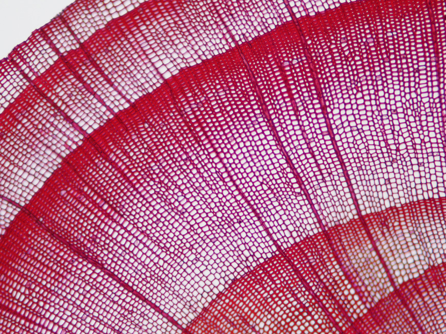
Cedar Annual Ring

Camellia Leaf Section

Leaf Stomata
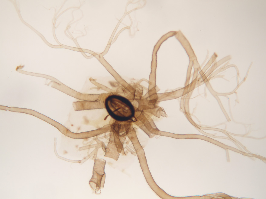
Insect Trachea

Pollen
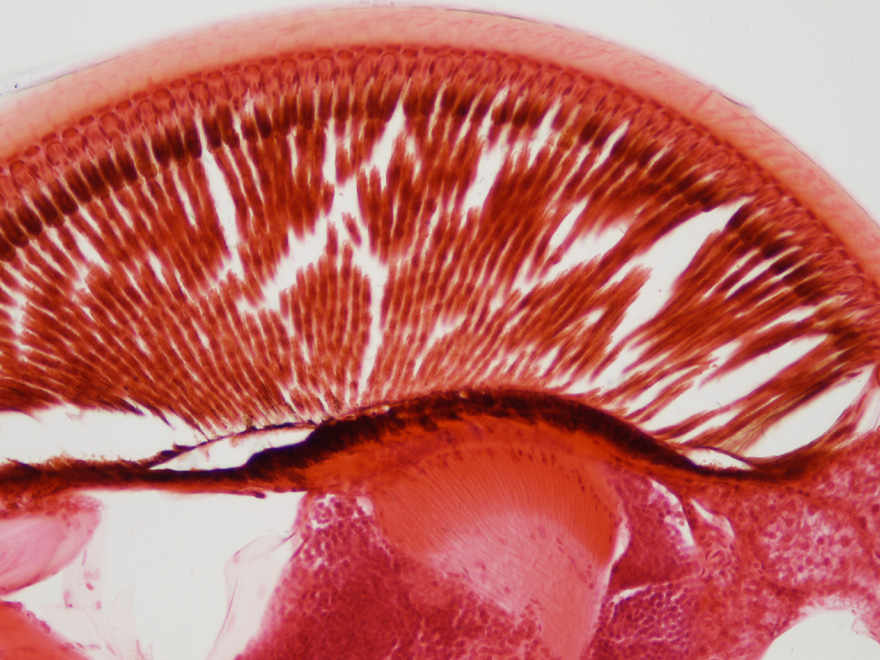
Insect Compound Eye

Pumpkin Stalk

Striated Muscle
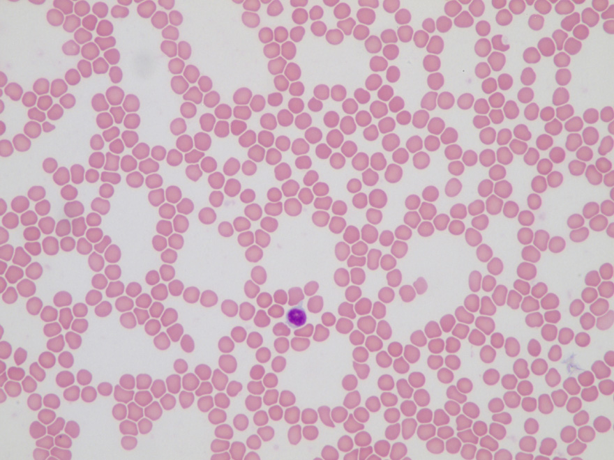
Blood Cell

Hibiscus Stem Section
Whole Organs Specimen - Sample Images

Cerebrum (40x)

Thyroid (20x)
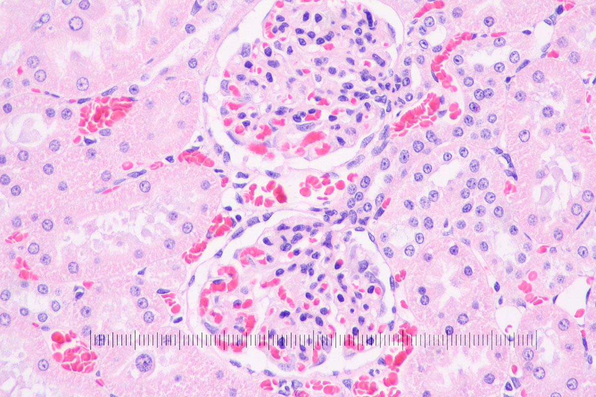
Kidney (40x)
These sample images are provided for reference. They are not a guarantee of identical image quality under every microscope, camera, illumination or observation condition.